With the new plastination collection at KHSU, medical students can encounter isolated organs and regions of the body.
Mastery of human anatomy is the cornerstone of reliable clinical diagnosis. With the arrival of plastinated human specimens to its anatomy lab, the Kansas College of Osteopathic Medicine (KansasCOM) is giving its students an edge: an opportunity to see anatomy as it truly exists in the body, variation and all.
Preserved in Germany through a process that transforms donor tissue into durable, odorless learning aids, plastinates allow students to explore the body in unprecedented detail to understand how form connects to function and how structure can reveal the roots of disease.
Director of anatomy laboratory Shannon Curran says she had long wanted to incorporate plastinates into KansasCOM’s non-cadaveric program, and the purchase was eventually made possible through Congressionally Directed Spending championed by Sen. Jerry Moran. The nearly $1 million in funding came through the Health Resources and Services Administration (HRSA) to expand simulation and anatomy programs at Kansas Health Science University (KHSU), the founding institution of KansasCOM, with the goal of improving health care for Kansans.
On a trip to the von Hagens Plastinarium last year, Curran saw the meticulous, two-phase plastination process firsthand. Expert dissectors replace water, blood and fats in donated human tissue with liquid polymers that harden, resulting in organs and systems for study with true-to-life color.
Curran says plastinates improve on traditional cadaveric anatomy instruction in many ways. Chiefly, using plastinated specimens means she has pristine, intact structures to reference every time she’s in the lab. With the two most common methods of anatomy study, standard dissection (by medical students) and prosection (where cadavers are dissected by instructors before lessons), there is a higher chance of severing important tissue and thus a lower chance of isolating key structures for replicated study.
“We have to remember that dissection is an inherently destructive process, no matter who’s doing it,” Curran says. “Preservation is the key in plastination. So, what we’re getting out of it is a perfect product. And we’re still getting all the key pieces, like variation and pathology because it’s a real human body. We’re getting it all.”
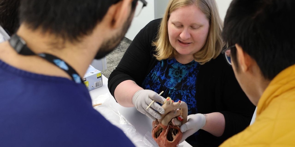
With the new plastination collection at KHSU, medical students can encounter isolated organs and regions of the body such as lungs, hearts, brains, arms, legs, hands, and even whole heads and necks.
Cameron Jeter, Ph.D., chair of anatomy and biomedical sciences, says the plastinates program “adds to our toolbox” resources that are supported by learning science, the study of how the human brain learns most effectively.
“There is no single learning style per person, as the idea that ‘I’m a kinesthetic learner,’ ‘I’m a visual learner,’ or ‘I’m an auditory learner’ is a brain myth. What is shown now to be the truth is multimodal learning, or stimulating as many of the human senses that we can,” Dr. Jeter says. “Bringing on plastinated specimens that are physically 3-D, manipulatable, and movable, where you can see and feel different dissections, enhances our program.”
By incorporating this multimodal approach across its curriculum, KansasCOM aligns its teaching methods with evidence-based strategies that maximize educational outcomes.
How Plastinates Improve Learning and Augment HoloAnatomy
Across all health professional schools, students generally study the same core anatomical structures. KHSU’s plastinated specimens were dissected and preserved specifically to view those structures from multiple axes, and Curran says she strives to bring in multiples of the same structures wherever possible to display realistic variation in age, sex, and body type.
Unlike wet cadavers, plastinates are pristinely dissected by experts before preservation. This allows both easier identification of anatomical structures and reliable assessment of student knowledge year after year via anatomy practicals, where students identify pinned, unlabeled structures. They also provide a complement to anatomical models, including virtual ones seen in HoloAnatomy, the mixed reality anatomy teaching tool using Microsoft HoloLens, which allows KansasCOM students to walk around and inside intangible structures.
KansasCOM student doctors in their first and second years will use the plastinates to supplement all anatomy lessons across courses under instructor supervision. Instructors will bring plastinates to afternoon lab sessions to reinforce the anatomy instruction from morning classes. This way, when students come to the anatomy lab, they are prepared to see physical representations of what they’ve learned. In years three and four, students can use them for review.
Dr. Jeter explained how the heart specimens, for example, display external tissues, miniscule internal structures, and how they interact.
“On the left side of the heart, you’re able to see the outside structures of the heart. But strategically, there’s been a hole dissected in the right side of the heart to look into the connection between the right atrium and the right ventricle and the valve that connects the two,” Dr. Jeter says. “Students can peek inside the chambers of the heart to visualize the blood flow, identify which chambers contain oxygenated or deoxygenated blood, and distinguish between bicuspid and tricuspid valves. This helps them grasp how these structures affect the heart’s ability to pump blood at various volumes and strengths.”
During his first introduction to KHSU’s plastinates in the cardiology unit, first-year medical student Justin Loh says he was able to compare the two hearts displayed and begin to appreciate differences not only in size but also in the number of arteries.
“One thing that I didn’t realize until Instructor Curran talked about it was some patients might have extra arteries, extra veins, or their ratios of this organ to that organ is a little different,” Loh says. “Veins, for example, have humongous variation to the point where it’s hard to teach the standardization of veins because there’s almost no standard. And then when you see the two hearts you notice one actually has four arteries when every single textbook shows three.”
In the anatomy lab, plastinated specimens handled by instructors are used in conjunction with HoloAnatomy, wherein students can virtually manipulate anatomy by magnifying and shrinking it and can walk around and inside virtual models.
“With the digital tools, you’re able to remove and add layers or labels, which is really nice. You couldn’t quite do that with the plastinates,” Loh says. “But the real organ is what gives you that emotional feeling and gives you the best perspective.”
Curran says the tools will be used in tandem, as many structures can be better understood after seeing them in multiple contexts.
“Sometimes the HoloLens makes things look too perfect. For example, you can visualize some tiny structures easily on HoloLens, but the plastinates show us that, in reality, they can be hard to find,” Curran says. “I’m thinking of the levator ani of the pelvis, so the pelvic floor. On HoloLens it shows you that you can differentiate those muscles really easily. But in reality you can’t. So, it’s a good comparison for students to have.”
How Plastinates Prepare Doctors for Patient Care
It took over a year from the time Curran placed the order for the plastinated specimens until the first shipment arrived in Wichita in a big wooden crate. The long timeline was a result of verification processes, international shipping, and ultimately the availability of ethically sourced donor bodies.
Fewer than 10 faculty members from around the world are invited each year to train at the world-famous von Hagens Plastinarium in Guben, Germany, known for preparing plastinates for medical and veterinary education, research, museum exhibits, and more. Thanks to KansasCOM’s order surpassing a specific threshold, Curran received the rare invitation for targeted training in how to maintain, protect, and repair the specimens, honoring the significant contributions of those who donated their bodies to science.
Dr. Jeter says that, for the viewer, interacting with real human parts like these elicits a greater emotional response—one of gratitude and appreciation—compared to virtual study.
“I realize these are fellow human donors who have been invested in the education I provide and the education my students receive, so I am grateful,” Dr. Jeter says. “Plastinates also evoke a sense of awe at the intricacies of the human body and its beauty, its innate design.”
She also mentioned the plastinates have infused new spirit into the anatomy faculty members as they think about the impact on future patients.
“Knowing anatomy is the first step to understanding the function and therefore the dysfunction of the human body. This is an essential continuum for the clinician,” Dr. Jeter says. “By understanding the intricate anatomy of how each finger or group of fingers is innervated, for example, you can listen to the patient’s complaint and quickly come to a differential diagnosis. If a patient’s thumb versus pinky finger goes numb, that informs the clinician which nerve is being pinched.”
Curran says if they are properly cared for, the plastinated specimens could last for decades.
“I am dead set on making sure that we handle them properly and we give them the utmost respect, so that they last 50 to 60 years,” Curran says. “I want them to outlast all of us here. I want them to be the longest lasting teachers here at KansasCOM.”
With careful stewardship, KHSU’s plastinates will continue shaping future physicians’ education. As the plastination library expands over time, these silent teachers will stand as a testament to the university’s commitment to modern medical education.
To help grow this invaluable resource and invest in Kansas medical students, contact the Office of Institutional Advancement. Your contribution can strengthen anatomy learning at KansasCOM and help to prepare tomorrow’s physicians to serve with deeper understanding and greater precision.